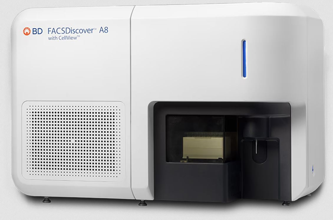
BD FACSDiscover A8 Cell Analyzer with BD CellView Image Technology
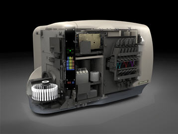
The BD FACSDiscover™ A8 Cell Analyzer with BD CellView Image Technology seamlessly integrates cutting-edge spectral flow cytometry with real-time imaging. By combining innovations such as BD SpectralFX™ Technology and BD CellView™ Image Technology into a single instrument, it offers unparalleled experimental power and simplified workflows to deliver reproducible results and single-cell insights that were previously undetectable. This versatile, robust and reliable instrument comes in a 4-laser configuration (488-637-405-561nm) with up to 56 fluorescence detectors. Imaging detectors together with fluorescence, scatter and light loss allow you to visualize events in real time and at high speed with 3-color fluorescence imaging off of the 488nm laser.
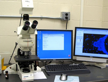
Location: EOHSI Building, Room 347